

· 4 min read
A pulse is a sentence, not a sum.
Heart rate counts your beats. The shape of each beat holds what the count leaves out.
Put two fingers on your wrist and count for a minute. You get a number, perhaps 62. It is a useful number. It is also a sum: sixty-two beats added together, with everything that made each one different left out.
Each beat has a shape. It climbs, crests, dips and swells again. Physicians were studying that contour in the nineteenth century (Millasseau et al. 2006), and traditional pulse readers attended to its qualities for centuries before. Read this way, a pulse is less a tally than a sentence.
What the light sees
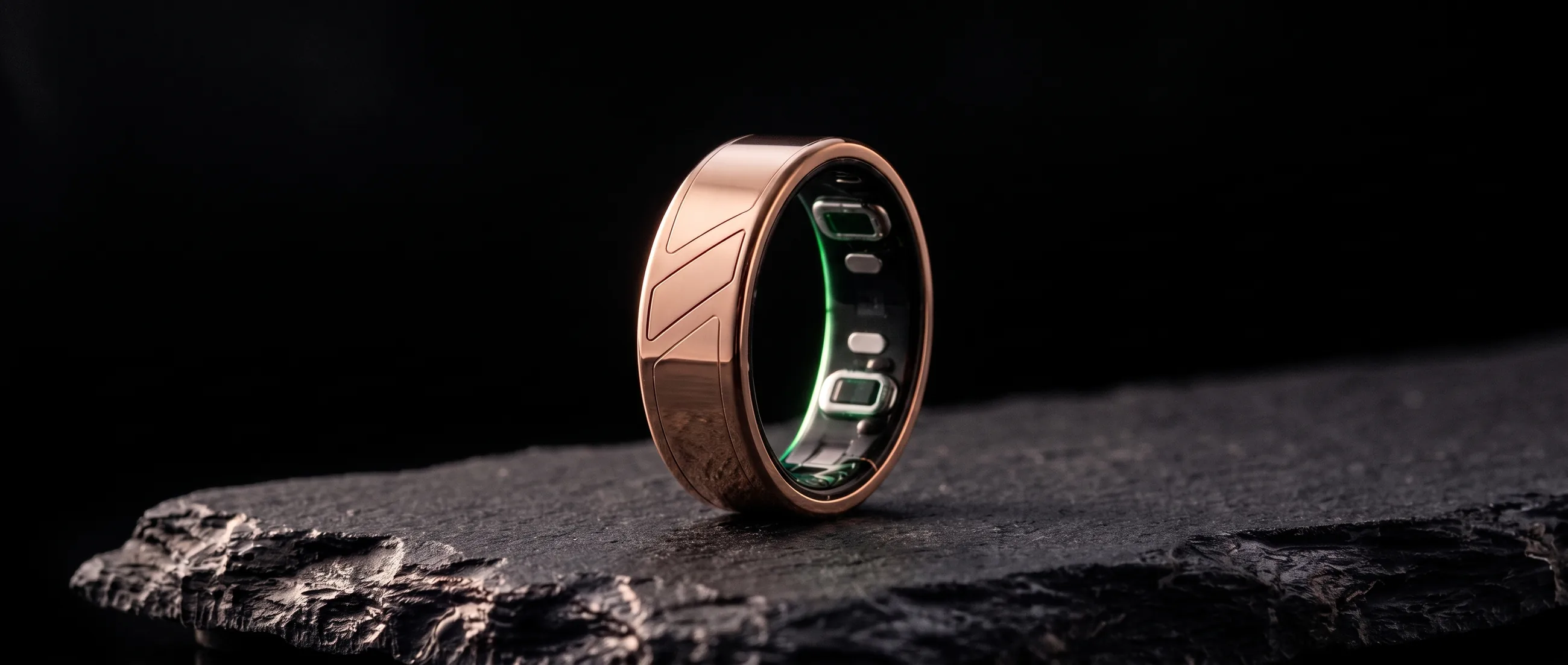
A ring can't feel your pulse the way a fingertip can. It shines light into the skin and measures what comes back. With each beat the small vessels under the sensor swell a little and ease again, and the light changes with them. The technique is called photoplethysmography. Its beat-by-beat signal rides on a slower tide shaped by breathing, the nervous system and body temperature (Allen 2007).
Draw a single beat of that signal, especially from a younger adult, and four parts stand out. You can watch them traced on our home page.
Four parts of a beat
- Rise. The upstroke (the systolic upstroke), as the wave from your heart arrives and the vessels under the ring fill. It carries the heart's push as your arteries deliver it. Researchers time it from foot to crest (Elgendi 2012).
- Peak. The fullest moment. Its height reflects how much the vessels swell with each beat, which has been related both to how much blood the heart sends and to how readily those vessels stretch (Elgendi 2012).
- Notch. A small dip on the way down, known as the dicrotic notch, where the heart's push ends and the valve at its outlet closes. It is usually clearest when arteries are supple (Elgendi 2012).
- Echo. A second, softer swell (the diastolic peak, or reflected wave). Much of it is thought to come from the part of each beat that travels down the aorta toward the lower body and is reflected back up to your finger (Millasseau et al. 2002; Elgendi 2012).
Why the shape matters
The echo keeps time. The stiffer the large arteries, the faster the wave travels and the sooner its reflection returns. In 87 adults, an index built from a person's height and the delay between the main wave and its echo correlated with a standard measure of aortic stiffness, taken between the neck and the groin (Millasseau et al. 2002).
The shape changes over a lifetime, too. With age the notch and the echo soften, until in later life they often can't be seen at all (Charlton et al. 2022). It also shifts as your vessels tighten or relax (Millasseau et al. 2006).
None of this survives the count. Two people can share a heart rate of 62 and have beats of quite different shapes. One number tells you how often. The shape tells you how.
It is the difference between counting the words in a sentence and reading it. Order matters. So do the pauses. Move the echo a little earlier and the beat says something different, though the count hasn't changed.
Researchers have been saying so for years. A review of fingertip pulse signals argued that the waveform deserves attention beyond oxygen readings and heart rate (Elgendi 2012). More recently, a model trained without labels on optical pulse recordings from about 141,000 smartwatch wearers learned representations that carried information about their demographics and health (Abbaspourazad et al. 2024).
The science is still unfolding. Where the optical signal comes from, both the beat and the slower tide beneath it, is not fully understood. Both are still generally accepted to offer useful information about the heart and circulation (Allen 2007). That is a reason to read carefully, and to compare you with you. None of these studies used Mere, and Mere doesn't measure arterial stiffness, blood pressure or any clinical marker.
Reading against your own normal
There is no single correct shape. Yours reflects your age, your build, your vessels and your night. A shape that is ordinary for one person can be a change for another. So Mere doesn't grade your pulse against a textbook. It learns yours.
Every night, the ring reads the shape of your pulse (its rise and peak, and its notch and echo where they show), alongside heart rate, heart rate variability, blood oxygen and sleep. Night is a good time to listen: your hand is mostly still, and movement blurs an optical pulse (Elgendi 2012). Mere learns what normal looks like for you over 14 nights. From then on, each night is read against it.
The pulse model behind those readings has been five years in development. What reaches you is plain English: a pattern such as Steady, Wired or Drained, drawn from your own nights rather than from an average.
It is a wellness reading, not a diagnosis, and it isn't meant to detect or monitor any medical condition. The ring notices. Your doctor decides.
Everybody is different. If you'd like to read your own pulse this way, join the founding list.
References
- Millasseau SC, Ritter JM, Takazawa K, Chowienczyk PJ. Contour analysis of the photoplethysmographic pulse measured at the finger. Journal of Hypertension. 2006;24(8):1449–1456. doi:10.1097/01.hjh.0000239277.05068.87
- Allen J. Photoplethysmography and its application in clinical physiological measurement. Physiological Measurement. 2007;28(3):R1–R39. doi:10.1088/0967-3334/28/3/R01
- Elgendi M. On the analysis of fingertip photoplethysmogram signals. Current Cardiology Reviews. 2012;8(1):14–25. doi:10.2174/157340312801215782
- Millasseau SC, Kelly RP, Ritter JM, Chowienczyk PJ. Determination of age-related increases in large artery stiffness by digital pulse contour analysis. Clinical Science. 2002;103(4):371–377. doi:10.1042/cs1030371
- Charlton PH, Paliakaitė B, Pilt K, et al. Assessing hemodynamics from the photoplethysmogram to gain insights into vascular age: a review from VascAgeNet. American Journal of Physiology – Heart and Circulatory Physiology. 2022;322(4):H493–H522. doi:10.1152/ajpheart.00392.2021
- Abbaspourazad S, Elachqar O, Miller AC, Emrani S, Nallasamy U, Shapiro I. Large-scale training of foundation models for wearable biosignals. International Conference on Learning Representations (ICLR). 2024. arXiv:2312.05409
Keep reading